


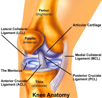
Articular cartilage is a firm, rubbery tissue that covers the ends of bones. Together with the thick synovial fluid within the joint, the cartilage forms a surface that allows low friction movement and assists as a shock absorbing tissue, distributing loads around the joint when walking or running.
When the cartilage is damaged, it does not repair itself effectively like other tissues. Consequently, injury can lead to increasing joint pain and reduction in joint movement and can also lead to degenerative change or osteoarthritis (slow degeneration of articular cartilage).
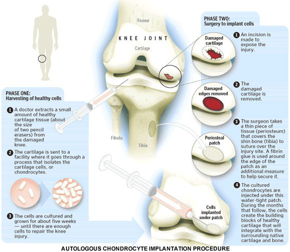
In certain situations, a treatment in which your own cells are implanted to repair articular cartilage damage can be performed. This procedure is known generically as a cartilage patch, or more specifically as Autologous Chondrocyte Implantation, or ACI. Autologous means that the implanted cells come from you, and the term chondrocyte is the name for the type of cartilage cells that repair the damaged area, known as a lesion.
ACI is typically reserved for patients who have sustained an injury resulting in full thickness or near full-thickness damage to the articular cartilage. It is not done when cartilage damage is due to degenerative arthritis (osteoarthritis) or when damage is extensive. More than one lesion can be treated with ACI in the same knee.
ACI is done in two separate procedures. In the first, small incisions (portals) are made around the joint. A scope is inserted into the knee. Saline solution flows through a tube (cannula) and into the knee to expand the joint and to improve visualization. The image is sent to a video monitor where the surgeon can see inside the joint. Surgical instruments go into an incision, and a small portion of healthy cartilage, called a biopsy, is removed from a non weight bearing region of the articular cartilage. After the biopsy, the surgical instruments are removed, and the first procedure is completed.
The location of the ACL in the knee joint makes it unlikely to heal when injured. Thus, reconstruction is the procedure of choice for restoring normal knee stability and avoiding further injury to the meniscus and/or cartilage. Most surgeons now favour reconstruction of the ACL using a graft from the patient's own leg to replace the torn ACL. There are two grafts commonly used to repair a torn ACL. One is a strip of the patellar tendon below the kneecap. The other is the hamstring tendongraft.
This injury has received a great deal of attention from orthopedic surgeons over the past 15 years, and very successful operations to reconstruct the torn ACL have been invented.
Your surgery will be performed by Dr. (Prof.) Anil Arora using an arthroscope (keyhole), a small fiber-optic video camera that is used to see and operate inside the joint. Only small incisions (portals) will be made during arthroscopy for this procedure. The surgery doesn't require Dr. (Prof.) Anil Arora to open the knee joint.
Cells collected from the biopsy are sent to a tissue engineering laboratory where the biopsy is processed, releasing the chondrocytes (articular cartilage cells) from the articular cartilage. These cells are placed in a nutrient rich medium and incubated which promotes cell growth. The culture process continues until an optimal cell number is attained. This can vary depending upon lesion size but normally ranges between 3 and 4 weeks.
The second operation is performed to implant the new cells into the damaged area. Dr. (Prof.) Anil Arora achieves this by sealing the damaged area with a waterproof cover and injecting the cells underneath the cover into this area. The cells attach to the bone and then begin the process of re-establishing the articular cartilage matrix. To ensure the implanted cells are protected, it is necessary to stay in hospital for a number of days to allow progressive joint mobilization.
A special rehabilitation programme needs to be followed following the implantation of cells. This involves bracing of the knee and a gradual return to full weight bearing. It is vital that the rehabilitation protocol given by Dr. Arora is followed closely to maximize the benefit of the operation.
This technique is mainly designed to repair small areas of cartilage damage resulting from accidental injury to the knee joint. It isn't yet proven for arthritis and would only be suitable for younger patients whose cartilage cells are more active.
The procedure requires mastery of difficult surgical techniques to achieve a good result. Dr. (Prof.) Anil Arora is one of the very few joint replacement surgeons in India to apply this highly skilled and ambitious treatment approach (ACI) to articular repair in young patients.